magazine_ Article
Brains in danger?
A lack of oxygen to the brain can cause serious damage, but the threat is difficult to measure. A young researcher is testing new approaches.
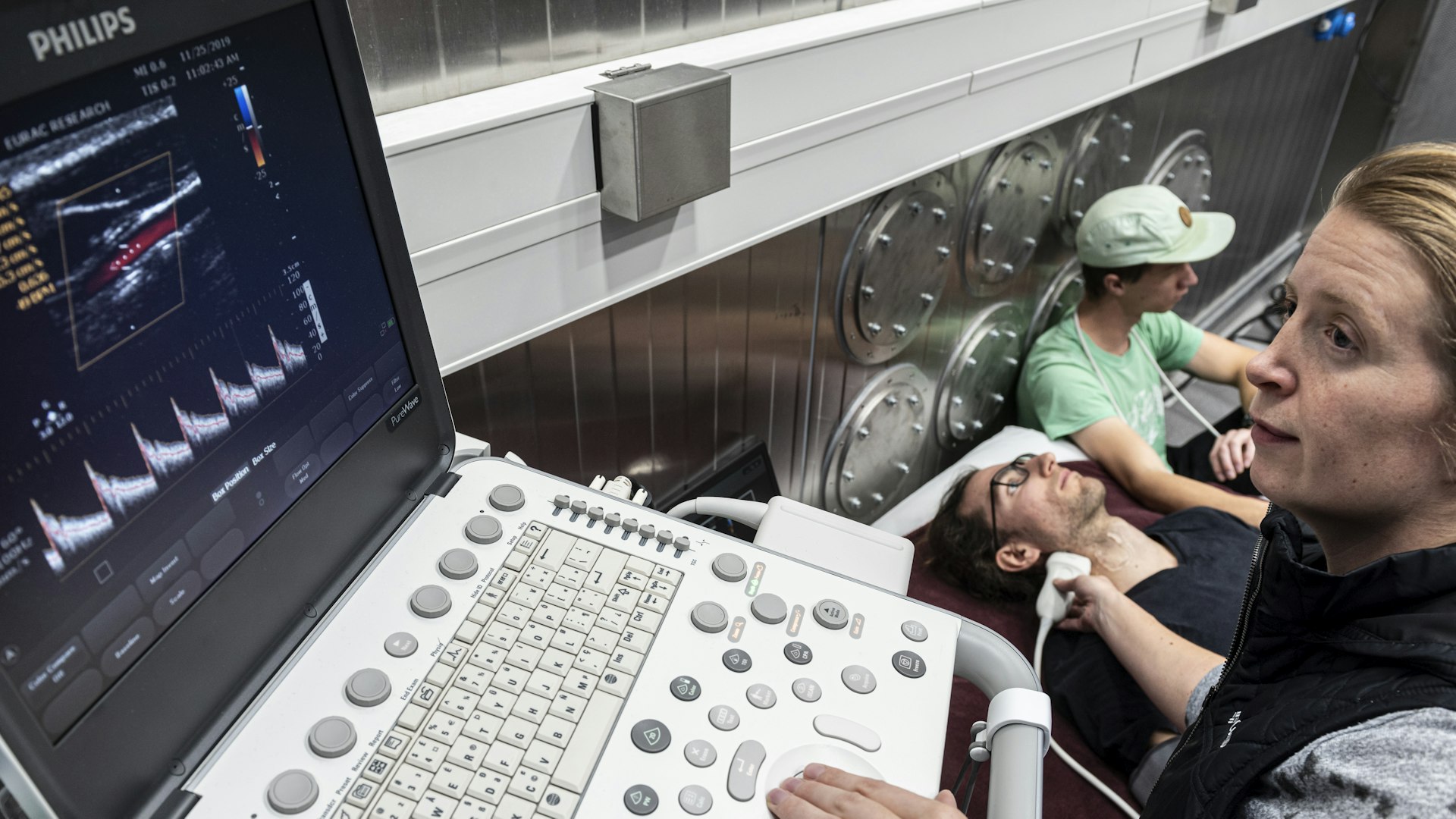
Ultrasound examination during a study in the terraXcube; the extreme climate simulator can be used under controlled conditions to investigate how the human body reacts to a lack of oxygen.
Credit: Eurac Research | Ivo Corrà
Oxygen deficiency in the brain can often cause permanent neuronal damage and occurs in half of all critically ill people in intensive care units. And as yet, there is still no simple and reliable indicator for the condition. Now Kai Riemer, an expert in ultrasound, is in the process of developing one. His method: seeing how healthy test subjects react to reduced oxygen in the terraXcube.
Kai Riemer moved to South Tyrol in June 2023, drawn to the region because of the extreme climate simulator terraXcube and its scientific implications as well as a passion for the mountains and the activities they provide. Riemer is a passionate photographer and hiker. Prior to this, he had spent many years as a researcher in London. Perhaps his time in the UK imbued him with British modesty, he apologizes, that he is “not so good at explaining” even after having adapted explanations of his research to the level of understanding of laypeople. One thing is without doubt: Riemer’s enthusiasm for ultrasound can be traced back to his research work in London. Ultrasound is a versatile, mobile, and inexpensive diagnostic tool, its smallest devices are the size of a cell phone, and as Riemer explains, used to have limited data capacity, however because of their immensely improved computers, no longer have any limitations. Riemer became an expert in blood flow imaging and super-resolution ultrasound imaging at Imperial College London, where he completed his doctorate and later went on to conduct research in various laboratories.
There are various ways to remedy the lack of oxygen. But in order to evaluate them, you first have to be able to measure the changes they bring about.
Kai Riemer
As a Marie Curie Fellow at the Institute of Mountain Emergency Medicine, he is now applying this expertise to a problem that affects many seriously ill people as well as mountaineers: oxygen deficiency in the brain. Specifically, he is working on methods to reliably quantify cerebral hypoxia, which can cause permanent neuronal damage and, in the worst case, brain death. His goal for the next two years is to study the condition in a non-invasive and simple way. The end goal: “What I actually want to attempt to do is intervene in the processes that trigger hypoxia and remedy this lack of oxygen. There are various ways to remedy the lack of oxygen. But in order to evaluate them, you first have to be able to measure the changes they bring about.”
If hypoxia occurs in healthy people because they ascend to altitudes where their body has less oxygen available, the remedy is simple: descend to lower altitudes. On the mountain to decide when this is advisable, it must be determined whether the victim has a headache or is dizzy. This is usually done with a questionnaire. “It’s qualitative, not quantitative, and is based purely on subjective assessment.” In intensive care units, on the other hand, doctors use an ultrasound scan of the optic nerve to assess the extent to which the lack of oxygen in the head has led to dilated blood vessels and thus increased intracranial pressure. However, this method is not objective either, explains Riemer: “It is actually undisputed in specialist literature that results vary greatly depending on who carries out the examination. Some doctors see no effect, others a big one.” Riemer suspects this lack of clarity could be related to the fact that these ultrasound images are two-dimensional, and testing this hypothesis is part of his research project. “To understand the mechanisms of the condition, the diameter of the nerve covering aka the optic nerve sheath is measured. It is therefore assumed the optic sheath nerve is a perfect cylinder. But what if it isn’t? Would a three-dimensional image provide a more accurate assessment? I don't know yet: it’s just a suggestion.”
He also has a second aim, not to improve an existing method, but to develop a completely new one: since what is used to draw conclusions about hypoxia in the head is not the optic nerve, but blood flow. Both are “surrogates” for hypoxia. This means they are parameters that are easier to measure, and which reliably indicate what you actually want to observe, in our case hypoxia – “it’s like the shadow of a tree in relation to the tree: if the shadow disappears, we can say with a fair degree of certainty that the tree is no longer there,” explains Riemer. And he uses a similarly vivid comparison to describe the propagation of the pulse waves on which his newly devised method is based: “If you make a garden hose wiggle with a movement, then the wave runs through the hose, and when it reaches the end, it is reflected back again. The propagation of this wave can be measured and calculated. In the case of the pulse wave, it changes depending on how rigid, elastic or how large the vessels are. And since the vessels in the head expand in hypoxia, I expect that this change can be recognized by the propagation of the pulse wave.” This is “very speculative”, he adds: “Well founded, but speculative nonetheless.” Ultrasound measurement would be much easier with this method and results could be obtained just by the ultrasound device being placed on the patient’s neck for a maximum of thirty seconds instead of having to take readings from their eye. If it works as Riemer envisages, the results will show very precisely the extent to which the patient’s head has already reacted to the hypoxia. “This could be expressed on a scale from zero to ten, or as a traffic light: green - yellow - red. Climbers could then be clearly told: your head is still fine; or if they’d be better off descending.”
Since the vessels in the head expand during hypoxia, I expect that this change can be recognized by the propogation of the pulse wave.
Kai Riemer
Riemer’s interest is not just in high-altitude medicine. Altitude only offers the opportunity to study the effect of oxygen deprivation on healthy people; people in intensive care usually suffer from so many things that it is impossible to filter out which changes are actually due to hypoxia. But there are also confounding factors in mountain studies too, namely the weather and the effort of the ascent. The extreme climate simulator, on the other hand, allows complete control: all undesirable influences can be eliminated and because of this, Riemer had the idea to conduct his research at the Institute of Mountain Medicine with the terraXcube. The fact that the unique laboratory is located in the small capital of a beautiful mountain region was also welcome. When Riemer applied for the placement, he had been hankering for the outdoors after spending years in the metropolis. Riemer had grown up in a green area of Berlin near many lakes and his whole family loves hiking and they used to visit the mountains twice a year. His passion for the outdoors took on a new dimension when he started taking ambitious landscape and nature photographs in the UK which can be seen on his website and Instagram account.
In the UK, he also organized “mini adventure tours”, such as climbing all the 3,000-foot-high mountains in North Wales, within 24 hours. “Everyone laughs about it here, but it’s a great landscape.” He organized the challenge in Wales as a fundraising event for the British Heart Foundation and is planning something similar in South Tyrol for the mountain rescue service.
For now, Riemer is not drawn back to London, although he continues to work with scientists from Imperial College – thin London living costs are too high. “This is generally a big problem today: salaries in research have not kept pace with inflation.” It is also due to this “reality of life” that he offers a sideline in professional drone videography which he really enjoys, and which also helps finance his expensive photography hobby. As a young researcher, you have to be resourceful. Or go into industry if you do that you just might be able to afford London, says Riemer. For him, it’s not a current option, after all he has an even bigger goal in mind.
Kai Riemer
Kai Riemer specializes in blood flow and super-resolution ultrasound imaging. He is currently a Marie Curie Fellow at Eurac Research’s Institute for Mountain Emergency Medicine. Previously, he worked at Imperial College London, where he also completed his PhD.
